

Industry-leading companies like Johnson & Johnson and Becton Dickinson are using the Carbon Digital Light Synthesis™ (Carbon DLS™) process and 3D printing materials to solve major healthcare challenges and inefficiencies. Alongside them are researchers with access to Carbon printers that are making breakthroughs themselves.
One of them is Alla Zamarayeva, PhD candidate in the Department of Electrical Engineering and Computer Sciences at the University of California, Berkeley. Zamarayeva leveraged Carbon Cyanate Ester 221 (CE 221) material to create a personalized, motion-minimizing surface coil that improves magnetic resonance imaging (MRI) (Figure 1).
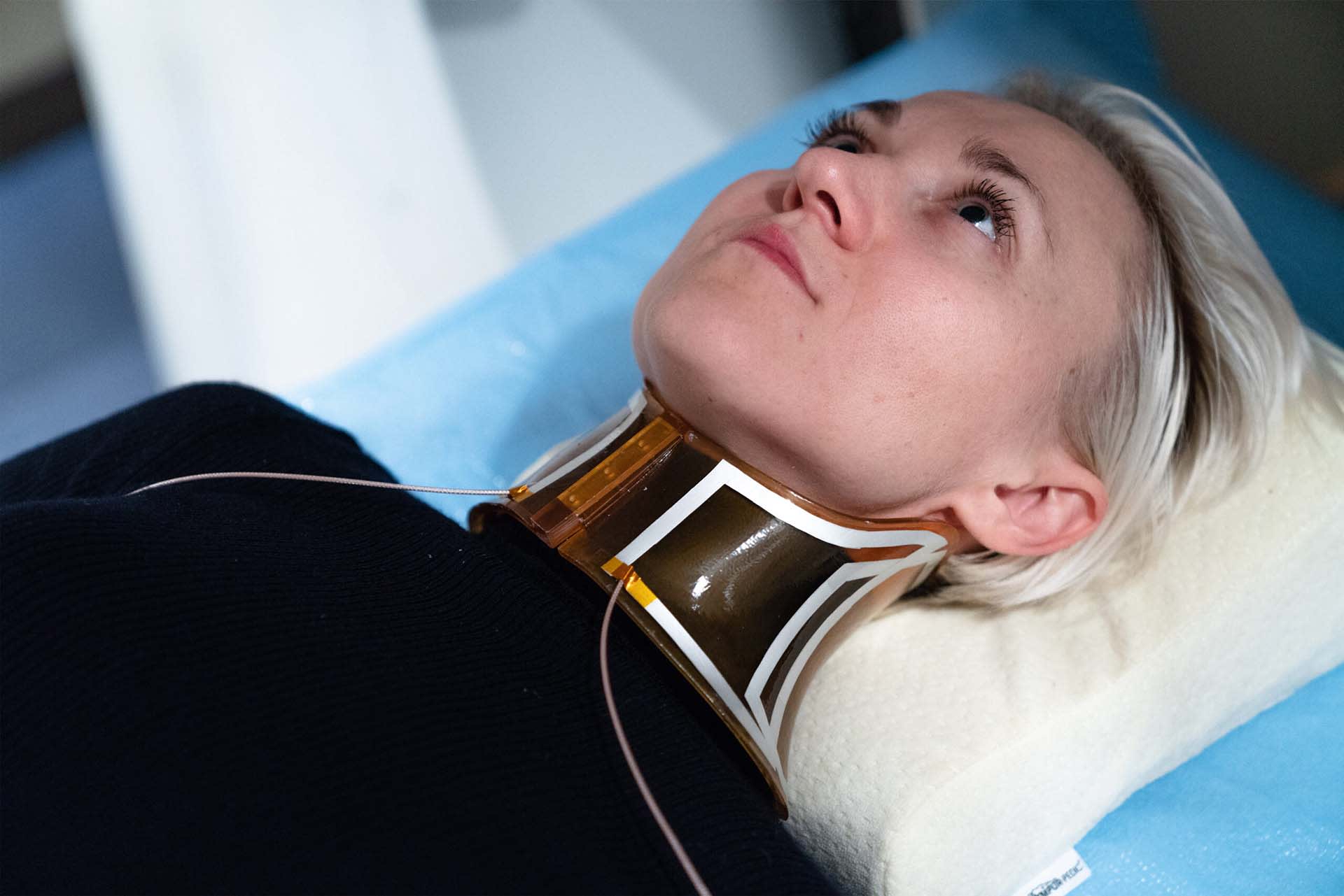
Improving MRIs can lead to significant advancements in clinical outcomes of diagnostics and next-generation therapeutic approaches (e.g. MRI-guided surgeries), which can offer better options for treating patients. However, there are two major technical challenges to obtaining high resolution MRIs: 1) patient movement during the exam and 2) inadequate signal-to-noise ratio.
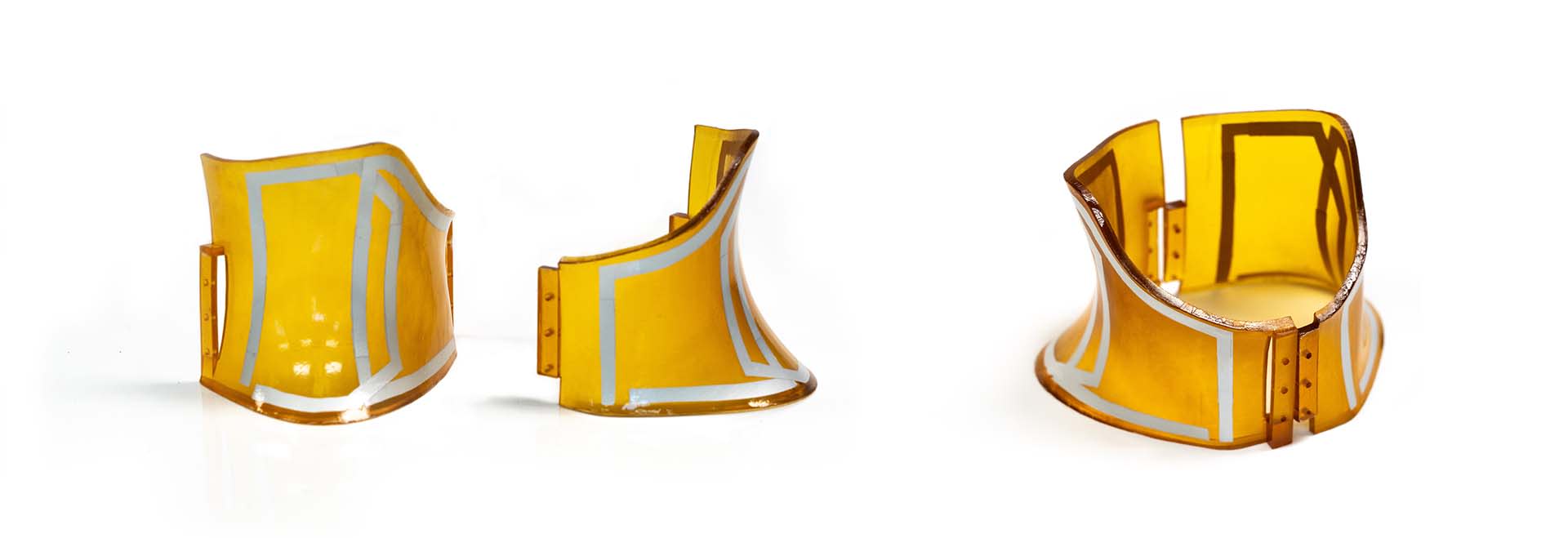
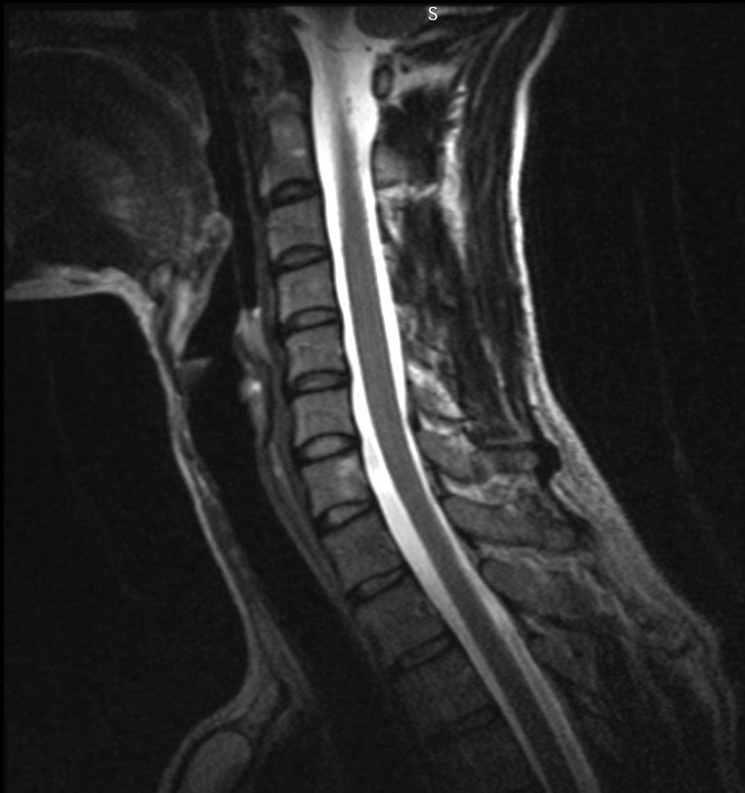
Under the guidance of Professor Ana Arias and Professor Miki Lustig, Zamarayeva created patient-specific coils for evaluating health conditions involving the neck that can, for example, detect spine lesions (Figure 2). The coils she created enhance the signal-to-noise ratio by bringing antennas as close as possible to the patient, resulting in higher resolution MRIs. With the unique combination of design freedom and high-temperature materials, the Carbon DLS™ process was able to make this innovation possible.
High-Temperature, Biocompatible 3D Printing Materials
MRI surface coils are radio-frequency (RF) antennas that pick up the MRI signal to produce the image. Coils are traditionally made in a single size and therefore do not fit each patient perfectly, resulting in reduced image quality. Researchers and designers have been searching for a technology capable of making a personalized solution for a surface coil: one that can be placed as close as possible to the patient and minimize motion.
When finding a material for a perfect fit, meeting the key mechanical properties of this application was crucial:
- Does not generate ghost MRI signals
- Low RF absorption to minimize signal attenuation
- Suitable for skin contact (biocompatible)
- Suitable for heating post-build for fabrication of the conductors on the surface of the printed part
So far, additive manufacturing technologies had only been used indirectly by printing templates that the substrate is formed on. The actual coils are then fabricated separately and attached to the substrate. Naturally, a more integrated print process would be preferable; but conventional 3D printing materials have not been able to withstand the high heat requirements for fabricating the surface coils directly on the surface. That’s where Carbon comes in.
High-temperature materials from Carbon offered the Berkeley team an ideal solution for the surface coils. The Carbon Cyanate Ester 3D printing material (Carbon CE 221) satisfied each of these requirements, including being able to withstand the high heat requirements needed to attach the coils directly on the surface without melting nor deforming like other plastic materials used for additive technologies would.
Creating the Digital File
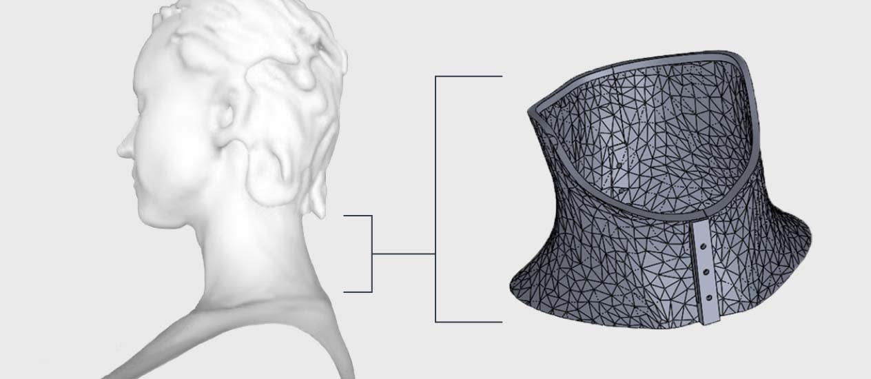
After finding a process and material that matched their application, the team began developing a prototype with photographic scans of a patient’s neck and shoulders created using a structural scanner and 3D modeling tool to generate a design for the surface coil (Figure 3).
Tailoring and Tracking by Patient
The Carbon DLS™ process enables end-use parts like the personalized surface coil to be designed and produced on-demand for a lot size of one, making truly patient-centric healthcare possible. To minimize any chances for error in printing a personalized device, the team incorporated a unique identifier into the print with ease using the integrated capability in Carbon software. This allows for careful and reliable tracking of the part through printing, post-processing, and delivery to the patient.
Metal to Complete the Surface Coil
Upon completion of the print, the team applied a metal coil to the surface of the CE 221 material. Using CE as the substrate enabled the collar to retain its shape in the oven while the conducting and insulating layers cured.
Improved Diagnosis with Sharper Image
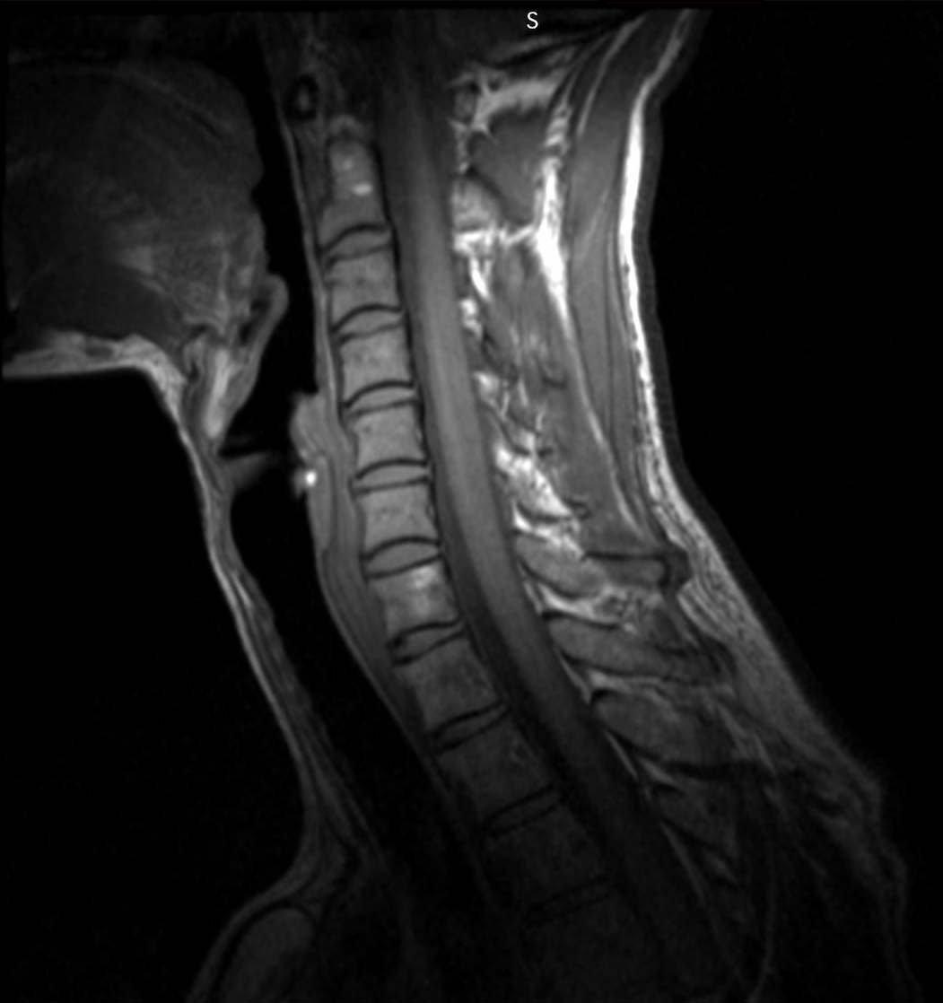
The researchers used the new digitally manufactured cervical collar and surface coil combination to acquire MRI images of test phantoms and a human subject. In the test done of MRI phantoms (a simple structure to ease analysis, not shown here), there was significant improvement of signal-to-noise ratios of the patient-specific coil images (Figure 4).
The excellent signal-to-noise ratio is noted by the high dynamic range of contrast in both T1 and T2 weighted images above. This enhanced resolution is critical for improved diagnosis and for more accurate procedure planning. Some improved diagnoses could include the visualization of tumors or the detection of plaque accumulation in the carotid artery, which can lead to a stroke.
Conclusion
Using the Carbon DLS™ process, Zamarayeva and researchers at UC Berkeley successfully digitally manufactured personalized devices and applied metal to high-temperature Carbon materials. This proof of concept work has implications beyond imaging. The ability to rapidly fabricate a personalized surface coil on complex surfaces capable of restricting motion can be coupled with the potential to introduce “portals” in the collar for directed energy delivery. This can lead to therapeutic uses of MRI such as MRI Directed High Intensity Focused Ultrasound (HIFU) for non-invasive surgical treatments.
We’re excited to help open up new possibilities for improving patients’ lives by enabling researchers like Zamarayeva with access to our software, technology, and materials.
If you’re interested in using Carbon technology and advanced materials to create personalized devices for better care, contact us at sales@carbon3d.com.
Note:
Material in this study was originally presented in a poster at the International Society for Magnetic Resonance in Medicine, June 16-18, 2018, Paris. France. (Zamarayeva A, Liu M, Corea J, et al. 3D sprayed MRI receive coils.)